

- Intake with the radiation oncologist
- Mould Room
- Simulation
- Treatment Planning
- Radiotherapy Treatment Delivery
- Check-ups during therapy
- Follow-up
Intake with the radiation oncologist
During the initial visits to the radiotherapy department, you will meet the radiation oncologist who will explain the treatment and its preparation – possibly you may have met her or him earlier. You will hear about side-effects that can occur during and shortly after treatment, but also in the years after treatment. The verbal explanation may be supplemented by written information. The doctor may examine your child and ask questions. The doctor will seek your consent for treatment, once you fully understand what is proposed. It can be helpful to prepare for the visit by writing down your questions for the doctor.
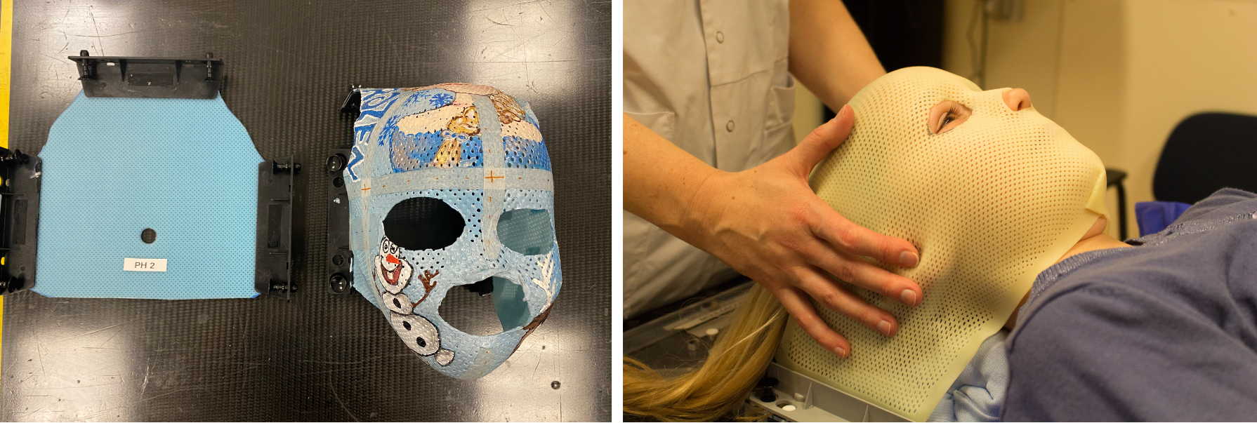
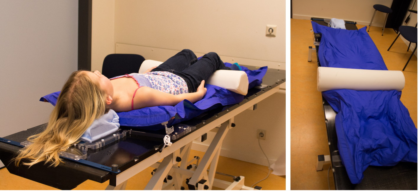
Mould Room
For the accuracy of treatment delivery it is imperative that the child remains still throughout the radiotherapy planning scan and treatment sessions. To help with the stability and reproducibility of patient positioning, a mould room appointment may be required prior to the acquisition of the radiotherapy planning scan. During this session, the radiographers will assess the child’s position and discuss what immobilisation equipment, if any, is required for treatment.
It is important to note that immobilisation equipment aids the reproducibility of patient positioning, enhancing the accuracy of the treatment delivery. The choice of equipment varies depending on the treatment site but also on local departmental choice.
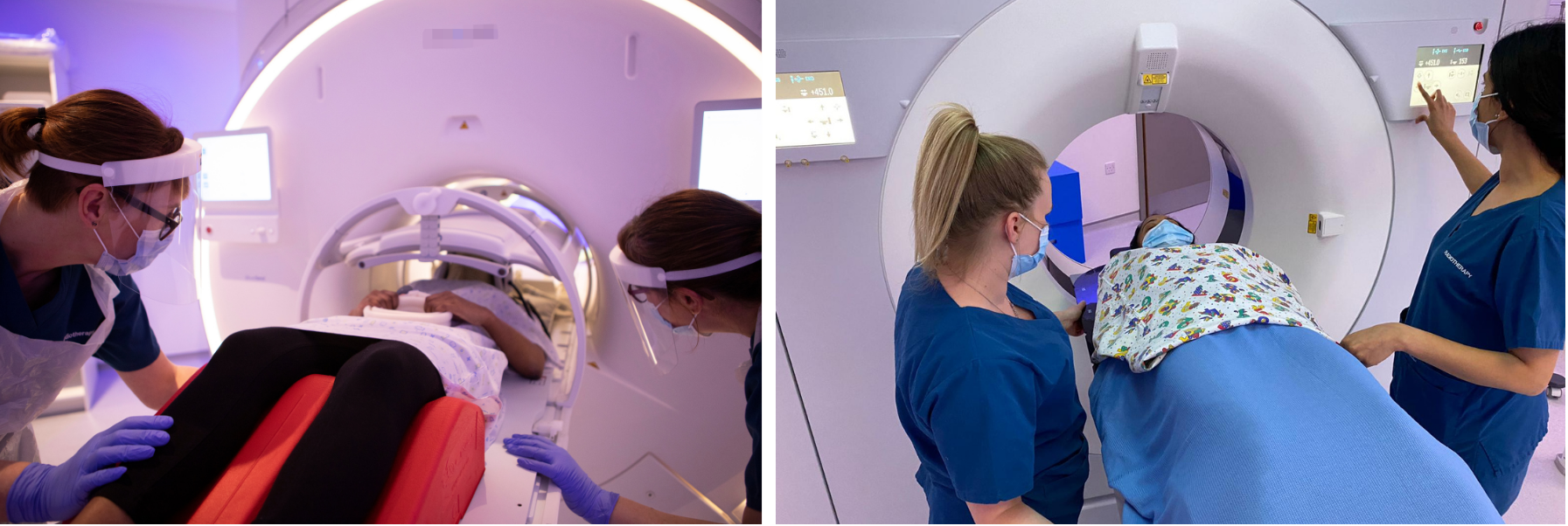
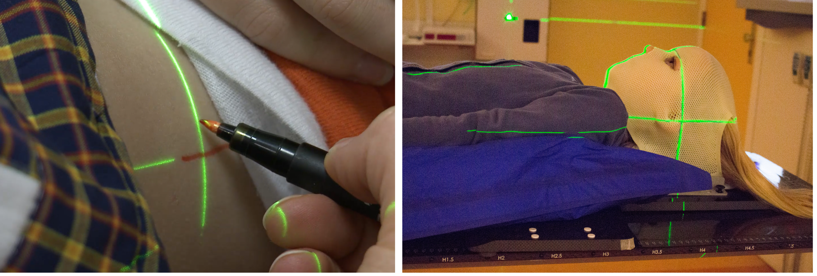
Simulation
Once positioning has been agreed the treatment planning phase can begin. This starts with a scan to show the precise location of the target region while in the treatment position. CT, PET-CT and MRI may be used for treatment planning purposes. The radiation oncologist will decide which scan is most suitable for each individual. The radiographers may take some time to re-assess the child’s positioning prior to the scan acquisition, while also using pens to draw marks on the skin. After the scan, the radiographers may then use a needle and ink to create a dot in the area previously marked using pens. Depending on the treatment area and the department’s procedures, the child may have 2-3 of these freckle-like dots which aid the accuracy and reproducibility of positioning for treatment. The radiation oncologist may also request the administration of intravenous contrast for the planning scan. Occasionally, the planning scan is repeated during treatment. This may, for example, be necessary in case of reduction of post-operative swelling or other anatomical changes.
Treatment Planning
After the treatment planning scan, the radiation oncologist outlines the area to be treated and works with the physics and dosimetry team to build an individualised radiotherapy treatment plan. This process can take up to a few weeks as the team decides how the radiation beams will be arranged, considering the avoidance of surrounding healthy tissues, whilst making sure radiation dose will be accurately delivered to the treatment area outlined by the radiation oncologist. Once the plan is complete it is then assessed through a series of checks before being finalised for treatment delivery.
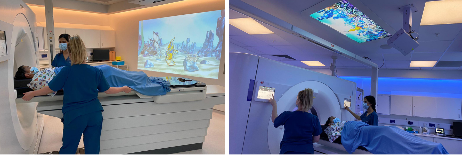
Radiotherapy Treatment Delivery
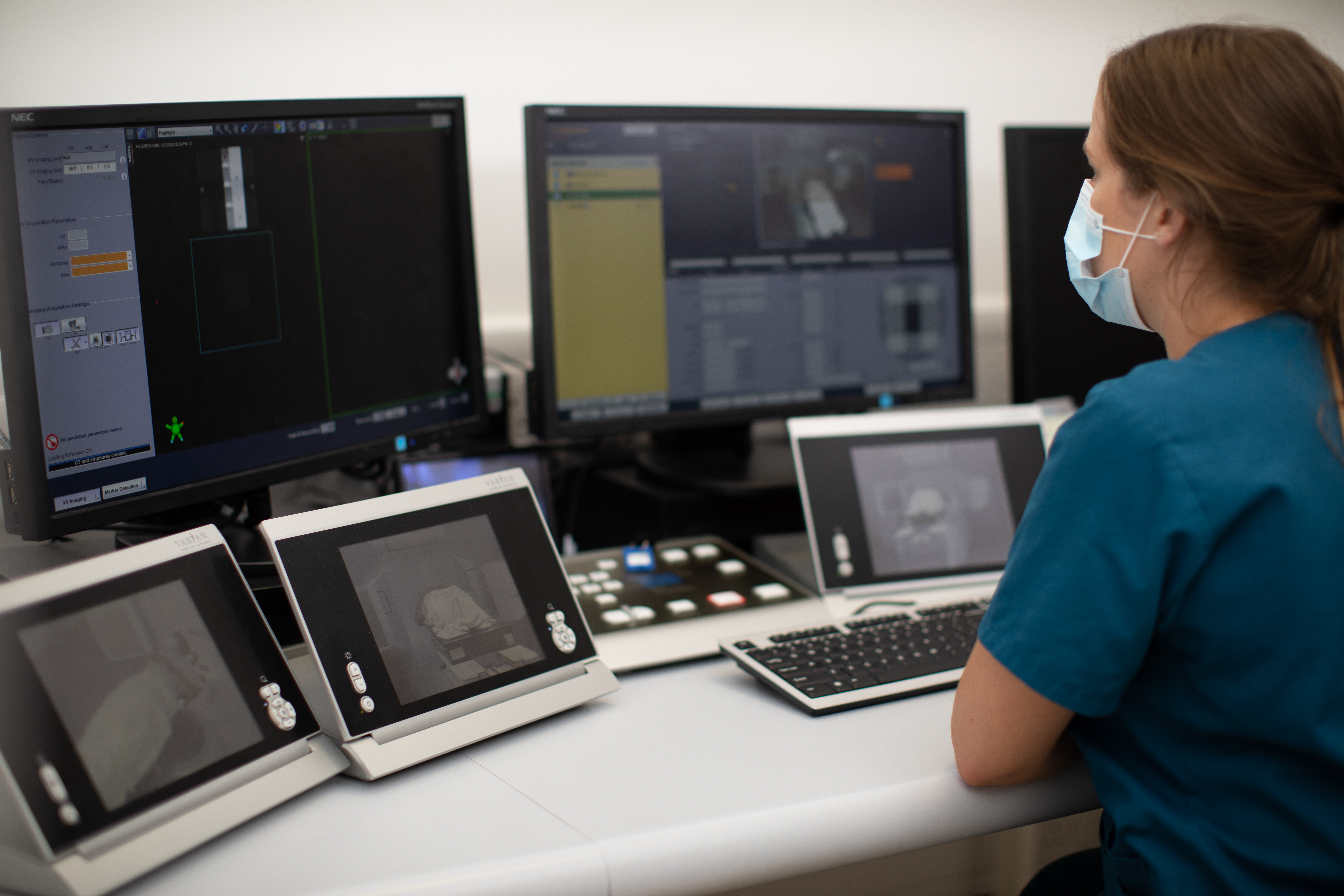
It is very important that the child’s positioning at the time of the planning scan acquisition is reproduced for treatment delivery. To do so ensures that the radiation beams are delivered with the same accuracy as planned by the radiation oncologist and the physics and dosimetry team. On the first day of treatment, the radiographers will take time to replicate the positioning, utilising the dots or pen marks on the patient’s skin and immobilisation device if required to align with millimeter thin laser lights that are projected on the skin. The radiographers will remind the child of the importance of remaining still during treatment set-up and delivery, while carrying out plan checks in the room.
Once the radiographers leave the room, they will take x-ray images of the treatment area to confirm the positioning is exactly the same as planned. If discrepancies are noted, the radiographers may return to the treatment room to re-position and re-image before proceeding to treatment delivery. During these checks and the treatment itself, the child is alone in the treatment room, it is not possible for anyone else to stay with the child.
Check-ups during therapy
During the treatment period, your child will regularly see the radiation oncologist or other team members to monitor the progress and check for side-effects. The radiation oncologist can prescribe medicines for side-effects if needed and discuss any questions or concerns about the treatment that you or your child may have. Urgent questions and physical complaints of your child can always be brought to the attention of any member of the radiotherapy team between doctor’s check-up appointments.
Follow-up
The radiation oncologist will monitor your child during the period of acute side-effects after radiotherapy. Depending on the center’s arrangements, the radiation oncologist will be involved in the long-term follow-up visits, or will be consulted by the pediatric oncologist if needed. During follow-up visits, patients will have diagnostic tests and/or scans to monitor the treatment response.